


When it comes to bone fractures, subtrochanteric fractures may not be as well-known as their more famous counterparts like wrist or hip fractures. However, subtrochanteric fractures are a serious orthopedic injury that can significantly impact an individual’s mobility and quality of life.To effectively manage and treat these fractures, orthopedic surgeons often rely on classification systems to guide decision-making.
One such classification system that plays a crucial role in understanding subtrochanteric fractures is the Seinsheimer Classification. In this blog post, we will explore what subtrochanteric fractures are, their causes, symptoms, treatment options, and the recovery process.
What is a Subtrochanteric Fracture?
A subtrochanteric fracture is a break in the femur bone, specifically located just below the hip joint and above the shaft of the femur. This region, known as the subtrochanteric region, plays a crucial role in supporting the body’s weight and facilitating leg movements. Therefore, fractures in this area can be particularly debilitating.
Causes of Subtrochanteric Fractures
Subtrochanteric fractures typically result from high-energy trauma, falls, or direct blows to the thigh. Common causes include:
Falls: Slipping, tripping, or falling from a significant height can generate enough force to fracture the subtrochanteric region.
Motor Vehicle Accidents: High-speed collisions can lead to subtrochanteric fractures, especially if the impact is concentrated on the thigh.
Sports Injuries: Athletes, particularly those involved in contact sports or activities that carry a risk of direct trauma to the thigh, may experience these fractures.
Pathological Conditions: In some cases, underlying conditions like osteoporosis or bone tumors can weaken the femur, making it more susceptible to fracture even with minimal force.
Seinsheimer Classification of subtrochanteric fracture
The Seinsheimer Classification is a system developed to categorise subtrochanteric fractures based on their anatomical characteristics. This classification system was introduced in 1978 by Dr. Fritz Seinsheimer, an orthopedic surgeon, to provide a comprehensive framework for describing these complex fractures.
The Seinsheimer Classification divides subtrochanteric fractures into four main types:
Type I: Simple Trochanteric Fracture
- This type involves a fracture line that extends through the greater trochanter but does not involve the femoral shaft itself.
- Type I fractures are typically the least severe of the four types and can often be treated with less invasive methods, such as a dynamic hip screw or intramedullary nailing.
Type II: Trochanteric with Basicervical Fracture
- Type II fractures extend through the greater trochanter and the cervical neck, causing a more significant disruption of the femoral anatomy.
- These fractures are generally more unstable and may require more extensive surgical intervention.
Type III: Comminuted Trochanteric Fracture
- Type III fractures involve a comminuted or fragmented greater trochanter.
- These fractures can be challenging to manage due to the lack of a stable bone fragment for fixation.
Type IV: Subtrochanteric Fracture
- Type IV fractures extend further down the femoral shaft, below the lesser trochanter.
- They are often considered the most severe subtrochanteric fractures, as they disrupt the integrity of the femoral shaft significantly.
Significance of Seinsheimer Classification
The Seinsheimer Classification is invaluable for several reasons:
- Treatment Guidance: It helps orthopedic surgeons determine the severity and complexity of the subtrochanteric fracture, allowing them to choose the most appropriate treatment strategy.
- Outcome Prediction: Different types of subtrochanteric fractures have different prognoses. By categorising the fracture, surgeons can offer patients more accurate information about their expected recovery and outcomes.
- Research and Education: The Seinsheimer Classification is an essential tool for research and education in orthopedics. It allows for standardised reporting and comparison of treatment outcomes across various institutions and studies.
The Seinsheimer Classification is a fundamental tool in the management of subtrochanteric fractures. It provides a structured way to categorise these complex injuries, guiding treatment decisions and offering valuable insights into prognosis.
As orthopedic knowledge and techniques continue to evolve, the Seinsheimer Classification remains a cornerstone in the field, ensuring that patients receive the best possible care for their subtrochanteric fractures.
Boyd and griffin Classification of subtrochanteric fracture
Subtrochanteric fractures represent a subset of hip fractures and are typically classified based on their location within the femur. These fractures occur approximately 5-7 cm below the lesser trochanter, where the femoral shaft transitions into the femoral neck.
Subtrochanteric fractures are often the result of high-energy trauma, such as motor vehicle accidents or falls from heights, but can also occur due to lower-energy mechanisms in individuals with weakened bones, like osteoporosis.
The Boyd and griffin Classification system was developed to provide orthopedic surgeons and clinicians with a comprehensive way to categorize subtrochanteric fractures, taking into account various factors that influence treatment decisions. This classification system has two main components:
Fracture Pattern: The Boyd and griffin Classification divides subtrochanteric fractures into four primary patterns:
- Type I: Simple transverse fractures.
- Type II: Complex fractures involving a combination of transverse and oblique components.
- Type III: Comminuted fractures with multiple fragments.
- Type IV: Reverse obliquity fractures, where the proximal fragment is in valgus and external rotation.
Fracture Stability: The classification system further stratifies these patterns based on fracture stability:
- A: Stable fractures.
- B: Unstable fractures with varus deformity (inward angulation) of the proximal fragment.
- C: Unstable fractures with valgus deformity (outward angulation) of the proximal fragment.
Clinical Significance
Understanding the Boyd and griffin Classification is crucial for several reasons:
- Treatment Planning: Different fracture patterns and stability categories require different treatment approaches. For instance, stable fractures (Type A) may be amenable to non-operative treatments, while unstable fractures (Types B and C) often require surgical intervention.
- Outcome Prediction: The classification system helps in predicting patient outcomes and complications. Unstable fractures tend to have a higher risk of complications, such as non-union, malunion, or implant failure.
- Research and Communication: The Boyd and griffin Classification facilitates effective communication among healthcare professionals and researchers, enabling them to compare treatment outcomes and conduct studies that can further improve fracture management strategies.
Symptoms of Subtrochanteric Fractures
Identifying the signs and symptoms of a subtrochanteric fracture is essential for prompt diagnosis and treatment. Common symptoms include:
- Severe Pain: Patients often experience intense pain in the thigh or groin area, making it difficult to bear weight on the affected leg.
- Swelling and Bruising: Swelling and bruising around the thigh may be evident due to tissue damage and bleeding.
- Inability to Move Leg: The injured leg may be immobilised, making it challenging to move or bend the hip or knee joint.
- Visible Deformity: In some cases, the fracture may cause a noticeable deformity or angulation of the thigh.

Treatment Options
The treatment of subtrochanteric fractures typically involves surgical intervention. Orthopedic surgeons use various techniques to stabilise the fracture and promote healing:
- Intramedullary Nailing: This is a common surgical procedure where a metal rod is inserted into the hollow center of the femur to hold the broken bone in place.
- External Fixation: In cases where intramedullary nailing may not be suitable, external fixators can be used to immobilise and align the fracture using pins and a frame outside the body.
- Plates and Screws: In some instances, metal plates and screws may be used to secure the fractured bones in place.
Recovery Process
The recovery process for subtrochanteric fractures can be lengthy and challenging. It involves a combination of the following:
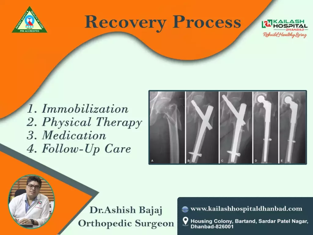
- Immobilisation: Patients may need to use crutches or a walker to avoid putting weight on the affected leg while it heals.
- Physical Therapy: Physical therapy is crucial to regain strength, flexibility, and mobility in the leg. It usually starts once the fracture shows signs of healing.
- Medication: Pain management and treatment for underlying conditions, such as osteoporosis, may be necessary to support recovery.
- Follow-Up Care: Regular follow-up appointments with the orthopedic surgeon are essential to monitor healing progress and make any necessary adjustments to the treatment plan.
Subtrochanteric fractures are significant orthopedic injuries that require timely diagnosis and appropriate treatment. While the road to recovery can be challenging, with the right medical care, physical therapy, and support, individuals can regain their mobility and quality of life. If you suspect a subtrochanteric fracture or experience symptoms, seek immediate medical attention to receive the best possible care and ensure a successful recovery.